Cervical screening guidelines have evolved faster than the equipment used to deliver them. Cytology timing, HPV co-testing intervals, and risk-based management algorithms have all been refreshed in the last five years — but in many practices, the colposcope itself is the same optical instrument from a decade ago. That gap matters, because the quality of the colposcopic exam shapes everything downstream: biopsy decisions, treatment selection, and the documentation trail that follows the patient for years.
In this guide, we’ll look at where video colposcopy fits in modern screening workflows, what features actually change clinical practice, and how the Edan C6AHD Video Colposcope stacks up against the everyday demands of a colposcopy clinic.
In this guide:
- Why video colposcopy is replacing traditional optical systems
- The clinical features that matter most at the exam table
- How the R-way™ evaluation system structures the four-stain workflow
- Specifications and workflow integration for the C6AHD
- When a video colposcope is the right purchase for your clinic
Why Video Colposcopy Is Replacing Traditional Optical Systems
Conventional optical colposcopes deliver the image to a single observer through binocular eyepieces. That worked for decades, but it limits three things modern clinics need: shared visualization for teaching and second opinions, digital documentation for the medical record, and image-based quality review.
Video colposcopes solve these problems by routing the exam through a high-resolution camera and display:
- Shared visualization — the entire care team, including residents and the patient when appropriate, can view the exam in real time
- Permanent documentation — every exam produces images and reports that live in the patient record, not just the colposcopist’s notes
- Quality review — colposcopic findings galleries allow side-by-side comparison across visits, which matters for surveillance of low-grade lesions
- Telemedicine and consultation — digital images can be shared with a specialist without requiring a second exam
The ASCCP risk-based management guidelines assume the colposcopist can accurately characterize lesion grade, location, and extent. Higher-quality imaging directly supports that characterization — particularly for distinguishing low-grade from high-grade disease at the squamocolumnar junction.
The Clinical Features That Matter Most
Not every feature on a colposcope spec sheet changes the exam. Three do.
1. LED cold lighting
Older halogen and xenon systems produce warm light that shifts tissue color and generates heat at the cervix. Modern LED cold lighting reproduces the native tissue color and stays comfortable for the patient during longer exams. The C6AHD uses a 40-LED array delivering ≥3,000 Lux of illumination — bright enough for clear visualization across the full magnification range without color cast.
2. Electronic green filter
Green-filter imaging is the cornerstone of vascular pattern recognition in colposcopy. Filtering out red wavelengths makes vessels stand out as dark lines against a pale background, which is essential for identifying mosaic patterns, punctation, and atypical vasculature — all features that drive the colposcopic impression. An electronic green filter (vs. a glass filter that must be physically swung into the optical path) lets the colposcopist toggle between white-light and green-light views without breaking eye contact with the exam.
3. Magnification range and depth of field
Useful colposcopy generally lives between 6x and 20x. The C6AHD covers 1-60x with real-time magnification display, which is more than most exams need but useful for documenting fine vascular detail or coaching a trainee through a specific finding. Operation distance of 200-300 mm at 6x matches the working space most colposcopists prefer.
The R-Way™ Evaluation System: Structuring the Four-Stain Workflow
Colposcopy is a four-step staining exam — saline, acetic acid, green filter, and Lugol’s iodine — and the colposcopist integrates findings across all four to produce an impression. The R-way™ evaluation system built into the C6AHD structures this integration into a memorable framework:
- R for Red (saline application) — reveals areas with rich blood circulation that reflect red under saline
- W for White (5% acetic acid) — reveals increased nuclear activity, which produces acetowhite epithelium
- A for Abnormal (green filter) — observes abnormal vascular features such as mosaic and punctation patterns
- Y for Yellow (Lugol’s iodine) — identifies iodine-negative areas that reflect yellow rather than mahogany
The system also includes an acetic acid and iodine reaction timer to ensure the colposcopist evaluates the cervix at the correct interval after stain application — a small detail that matters because acetowhitening evolves over time, and reading it too early or too late distorts the impression.
This structured approach aligns with the ASCCP Colposcopy Standards, which emphasize systematic documentation of lesion characteristics rather than gestalt impressions alone.
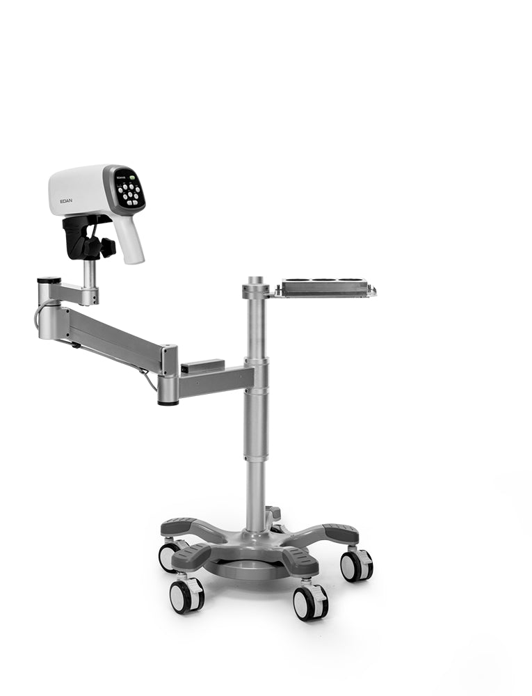
C6AHD Specifications at a Glance
| Specification | C6AHD Video Colposcope |
|---|---|
| Imaging | 2.13 MP camera, HD signal, ≥900 TVL system resolution |
| Magnification | 1-60x, real-time display |
| Operation distance | 200-300 mm at 6x |
| Field of view | ≥100 mm at 3x; ≥15 mm at 18x |
| Illumination | 40-LED cold lighting, ≥3,000 Lux |
| Focus | Manual and auto |
| Filters | Electronic green filter |
| Software | DICOM 3.0 compatible, R-way™ evaluation, findings gallery, PDF report export |
| Mounting options | Vertical stand, swing-arm stand, or MT-806 trolley |
| Operation | Remote capture control, one-hand keyboard layout |
Takeaway: For practices replacing an aging optical colposcope or building a new colposcopy clinic from scratch, the C6AHD’s combination of HD imaging, electronic green filter, structured R-way evaluation, and DICOM-compatible patient management software covers the full exam-to-report workflow without requiring add-on software.
Workflow Integration: From Exam Room to EMR
The clinical value of a video colposcope only materializes if the captured images and reports get to the right place. The C6AHD’s patient management software supports the full chain:
- Enter patient information at the start of the exam
- Capture and tag images at each staining phase
- Analyze findings in the R-way framework with side-by-side comparison
- Export the final report as PDF for the medical record or referring provider
- Print directly from the system when a hard copy is needed for documentation or patient handout
DICOM 3.0 support matters for clinics integrating with hospital PACS or EMR systems — it means the C6AHD’s images don’t live in a separate, isolated database. For high-volume practices, this is the difference between a colposcope that fits into existing workflows and one that creates a parallel documentation track.
When a Video Colposcope Is the Right Purchase
A modern video system isn’t necessary for every practice, but it’s the right call when:
- Your existing colposcope predates current ASCCP standards (typically pre-2017)
- You’re a teaching practice where residents need to view the exam alongside the attending
- You document a high volume of biopsies and need image-based records for surveillance
- You’re integrating colposcopy into a multi-specialty clinic and need DICOM-compatible output
- You manage low-grade lesions over multiple visits and need consistent image comparison
The NCI cervical screening guidelines for healthcare professionals and the CDC cervical cancer screening recommendations both reinforce the shift toward risk-based, longitudinal management — and longitudinal management depends on consistent, comparable documentation across visits.
Browse Minerva’s colposcopy instruments and accessories or request a quote for the C6AHD Video Colposcope for pricing, mounting configuration, and software setup.
This article is for informational purposes for healthcare professionals. It does not constitute medical advice or replace clinical judgment. Always follow your institution’s protocols and the manufacturer’s instructions for use. Refer to current ASCCP guidelines and ACOG practice advisories for cervical screening management.


